|
Problems after ankle fractures
|
The ankle
joint is a unique structure which depends on perfect alignment
of the bones for normal function. Although the ankle moves
in an up and down direction (called dorsiflexion and plantarflexion),
there is very subtle movement with twisting, called rotation.
In order for the ankle to work efficiently, the bones need
to line up perfectly in the socket, which we call the mortise.
If you look at the pictures below, it is easy to see the difference
between a perfect ankle anatomy, and one which is very deformed
following a fracture. |
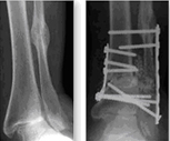
| |
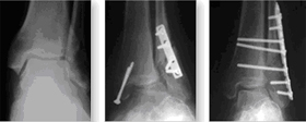
On the left is a picture
of a normal ankle XR. The shadow in the middle of the ankle
joint is the cartilage space, which does not show up on an
XR. Note on the middle XR the changes in the position of the
ankle after a fracture which was inadequately fixed with a
plate and screws. This is a serious problem and is associated
with terrible deformity. This was well reconstructed by re-breaking
the ankle, and fixing it again with a plate and screws as
can be seen on the right hand XR. |
| |
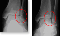
On the left again
is the normal XR of the ankle. It is easy to see the overlapping
shadow on the outside of the ankle which indicates the normal
bone structure. The overlap is highlighted in the circle.
|
| |
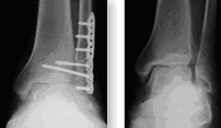
On the left hand XR
you can see again the space between the talus and the fibula
which is abnormal. This occurred after a fracture of the fibula
(called a high fibula fracture, and which cannot be seen on
this XR). If the ankle is left in this position with the fibula
shifted outward, arthritis of the ankle will inevitably occur.
In order to correct this, the fibula had to be broken again
and then fixed with a plate and screws. You can see that the
space between the talus and the fibula has now been corrected. |
|
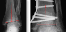
There are times when
the deformity which occurs after an ankle fracture can be
subtle, but still cause severe problems leading to arthritis.
It has been demonstrated scientifically that even very slight
shifts of the ankle of 2mm can lead to the development of
arthritis. In the XR on the left, the fibula was fractured,
but not treated with surgery. This is a difficult problem,
since the ankle no longer lines up correctly, and was treated
by making a large cut in the tibia bone, and removing a wedge
of bone from the tibia to line it up correctly. This can be
seen on the red lines which should be exactly perpendicular
to each other as on the right hand XR following the corrective
surgery. |
|
There are times when
the deformity is really severe as in this patient with rheumatoid
arthritis. This patient suffered from a stress fracture of
the fibula, and the ankle then collapsed, and the foot became
severely flat. This had to be reconstructed by cutting both
the tibia and the fibula (called an osteotomy) and then by
inserting a large specialized bone graft in both the tibia
and the fibula to re-position the ankle correctly. |
| |